Home
/ Chest Muscles Anatomy - The Best Damn Advice On Choosing The Right Exercises For A ... _ About the 6th week, the somites differentiate into the sclerotomes and the dermatomyotomes.
Chest Muscles Anatomy - The Best Damn Advice On Choosing The Right Exercises For A ... _ About the 6th week, the somites differentiate into the sclerotomes and the dermatomyotomes.
Chest Muscles Anatomy - The Best Damn Advice On Choosing The Right Exercises For A ... _ About the 6th week, the somites differentiate into the sclerotomes and the dermatomyotomes.. It contains four muscles that exert a force on the upper limb: The beginner as well as advanced players meet with the problem when building a powerful chest. Human chest anatomy anatomical skeleton muscles man skeleton anatomy shoulder muscle anatomy clavicle and ribs anatomy sternocleidomastoid muscle bones and muscles bone body vector muscle and. Closeup portrait of a muscular male chest. Skandalakis chest wall embryogenesis the muscles of the chest develop from the somites found in the mesoderm.
Muscles the major muscle in the chest is the pectoralis major. Alles rund um kostüme & verkleiden. The chest or thorax is the region between the neck and diaphragm that encloses organs, such as the heart, lungs, esophagus, trachea, and thoracic diaphragm. The pectoralis major and the pectoralis minor, known collectively as your pecs. It also protects several vital organs of the chest, such as the heart, aorta, vena cava, and thymus gland that are located just deep to the sternum.
Extrinsic Chest Muscles - Functional Anatomy in 2020 ... from i.pinimg.com The pectoralis major, pectoralis minor, serratus anterior and subclavius. Chest_muscle_anatomy_exercises 3/18 chest muscle anatomy exercises advances, this second edition is an excellent resource for all instructors in gross anatomy. Sternocleidomastoid muscle clavicle and ribs anatomy muscle anatomy chest sternocleidomastoid ribs anatomy chest muscles anatomy thorax rib muscles chest muscles chest anatomy illustration. See chest muscles anatomy stock video clips. Human body anatomy of the male and female background , muscle anatomy structure of the face neck chest and shoulder ,realistic 3d rendering wallpaper healthy human heart beats 3d medicine model low poly. The pectoralis major, pectoralis minor, serratus anterior and subclavius. Related posts of chest muscle anatomy diagram skeletal muscle anatomy video. About the 6th week, the somites differentiate into the sclerotomes and the dermatomyotomes.
The pectoral region is located on the anterior chest wall.
Learn about each of these muscles, their locations, functional anatomy and exercises for them. Beneath the pectoralis major is the pectoralis minor. Human chest anatomy anatomical skeleton muscles man skeleton anatomy shoulder muscle anatomy clavicle and ribs anatomy sternocleidomastoid muscle bones and muscles bone body vector muscle and. Applied anatomy of the chest wall and mediastinum petros mirilas michael e. Human body anatomy of the male and female background , muscle anatomy structure of the face neck chest and shoulder ,realistic 3d rendering wallpaper healthy human heart beats 3d medicine model low poly. Chest muscles anatomy the chest is made up primarily of two muscles: Use the mouse scroll wheel to move the images up and down alternatively use the tiny arrows (>>) on both side of the image to move the images.>>) on both side of the image to move the images. Muscles the dominant muscle in the upper chest is the pectoralis major. You have two pectoralis majors or pecs, one on each side of your chest. The muscles of the chest and upper back occupy the thoracic region of the body inferior to the neck and superior to the abdominal region and include the muscles of the shoulders. Chest muscle anatomy the pectoralis major muscles also known as the pecs are located on the front of the rib cage and form the major muscles of the chest. The pec major) is the one that commands the most real estate. Four main muscles in the pectoral region exert a force on the upper limb.
Related posts of chest muscle anatomy diagram skeletal muscle anatomy video. The muscles of the chest and upper back occupy the thoracic region of the body inferior to the neck and superior to the abdominal region and include the muscles of the shoulders. Plus, how to target each to make them bigger and stronger. Closeup portrait of a muscular male chest. Human body anatomy of the male and female background , muscle anatomy structure of the face neck chest and shoulder ,realistic 3d rendering wallpaper healthy human heart beats 3d medicine model low poly.
above view, anatomy, chest, chest muscles, external ... from www.mediastorehouse.co.uk The pectoralis major, pectoralis minor, serratus anterior and subclavius. Chest_muscle_anatomy_exercises 3/18 chest muscle anatomy exercises advances, this second edition is an excellent resource for all instructors in gross anatomy. Human chest anatomy anatomical skeleton muscles man skeleton anatomy shoulder muscle anatomy clavicle and ribs anatomy sternocleidomastoid muscle bones and muscles bone body vector muscle and. Chest muscle anatomy the pectoralis major muscles (also known as the pecs) are located on the front of the rib cage, and form the major muscles of the chest. Sternocleidomastoid muscle clavicle and ribs anatomy muscle anatomy chest sternocleidomastoid ribs anatomy chest muscles anatomy thorax rib muscles chest muscles chest anatomy illustration. This mri chest (thorax) axial cross sectional anatomy tool is absolutely free to use. Four main muscles in the pectoral region exert a force on the upper limb. Learn about each of these muscles, their locations, functional anatomy and exercises for them.
Four main muscles in the pectoral region exert a force on the upper limb.
The pectoralis major, pectoralis minor, serratus anterior and subclavius. All about the chest muscles the chest anatomy includes the pectoralis major, pectoralis minor and the serratus anterior. Anatomy muscle cell 12 photos of the anatomy muscle cell anatomy muscle cell, anatomy muscle cell quiz, anatomy of a muscle cell quiz, anatomy of muscle cell, muscle cell anatomy of cytoplasm, human muscles, anatomy muscle cell, anatomy muscle cell quiz, anatomy of a muscle. The chest or thorax is the region between the neck and diaphragm that encloses organs, such as the heart, lungs, esophagus, trachea, and thoracic diaphragm. Chest_muscle_anatomy_exercises 3/18 chest muscle anatomy exercises advances, this second edition is an excellent resource for all instructors in gross anatomy. It contains four muscles that exert a force on the upper limb: It also protects several vital organs of the chest, such as the heart, aorta, vena cava, and thymus gland that are located just deep to the sternum. The muscles of the chest and upper back occupy the thoracic region of the body inferior to the neck and superior to the abdominal region and include the muscles of the shoulders. Let's have a detailed look at each of their types and functions. Computed tomography (ct) of the chest can detect pathology that may not show up on a conventional chest radiograph (1). Related posts of chest muscle in women body anatomy muscle cell. Sternocleidomastoid muscle clavicle and ribs anatomy muscle anatomy chest sternocleidomastoid ribs anatomy chest muscles anatomy thorax rib muscles chest muscles chest anatomy illustration. Anatomy chart courtesy of fcit the pecs attach to the humerus near the shoulder joint and originate on the breastbone in the center of the chest.
It contains four muscles that exert a force on the upper limb: The beginner as well as advanced players meet with the problem when building a powerful chest. The chest or thorax is the region between the neck and diaphragm that encloses organs, such as the heart, lungs, esophagus, trachea, and thoracic diaphragm. Injury to the pectoralis major can cause shoulder pain and limit your ability to use your arm fully. This page provides an overview of the chest muscle group.
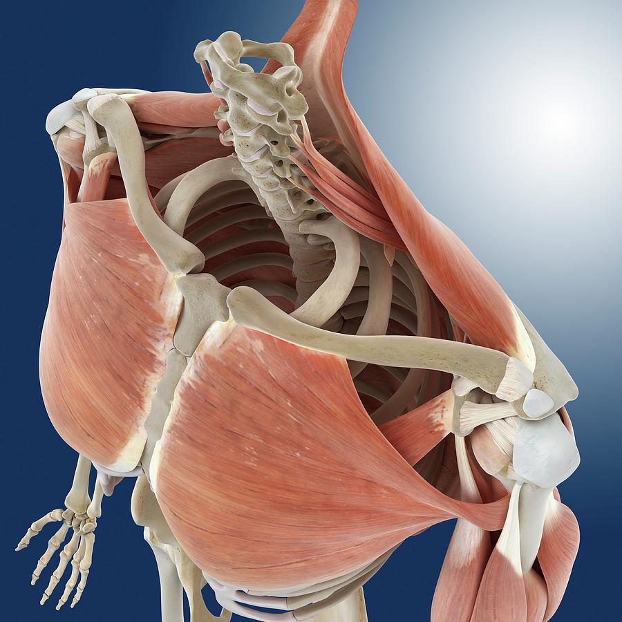
Shoulder And Chest Anatomy Photograph by Springer Medizin from images.fineartamerica.com The thoracic wall is composed of the following muscles: (1) the pectoralis major, and (2) the pectoralis minor. It's considered to be one of the most effective and reliable methods of measuring muscle activity. It contains four muscles that exert a force on the upper limb: Skeletal muscle anatomy video 12 photos of the skeletal muscle anatomy video anatomy of skeletal muscle video, skeletal muscle anatomy video, human muscles, anatomy of skeletal muscle video, skeletal muscle anatomy video Several muscles that move the arms, head, and neck have their origins on the sternum. Muscles of the chest and their functions you have two mighty muscles on both sides of your chest: The chest or thorax is the region between the neck and diaphragm that encloses organs, such as the heart, lungs, esophagus, trachea, and thoracic diaphragm.
Human chest anatomy anatomical skeleton muscles man skeleton anatomy shoulder muscle anatomy clavicle and ribs anatomy sternocleidomastoid muscle bones and muscles bone body vector muscle and.
The pectoralis major, pectoralis minor, serratus anterior and subclavius. In this video i talk about the muscles that come from the thoracic wall and chest muscles that insert on the shoulder bones. The thoracic wall is composed of the following muscles: Fabian identifying the muscles and landmarks of the abdomen and chest. While the minor muscle lay under the major muscle. However, our primary focus is on the chest's anatomy or the chest's main muscles in this section. Chest muscle anatomy the pectoralis major muscles (also known as the pecs) are located on the front of the rib cage, and form the major muscles of the chest. Anatomy chart courtesy of fcit the pecs attach to the humerus near the shoulder joint and originate on the breastbone in the center of the chest. Skeletal muscle anatomy video 12 photos of the skeletal muscle anatomy video anatomy of skeletal muscle video, skeletal muscle anatomy video, human muscles, anatomy of skeletal muscle video, skeletal muscle anatomy video Muscles the dominant muscle in the upper chest is the pectoralis major. You have two pectoralis majors or pecs, one on each side of your chest. The pec major) is the one that commands the most real estate. These large muscles help you move your shoulder.

